Explainer · Skin
What a multispectral skin scan actually measures
White, ultraviolet, and cross-polarized light each read a different depth of your skin. Eight measures come out; here is what each one means.
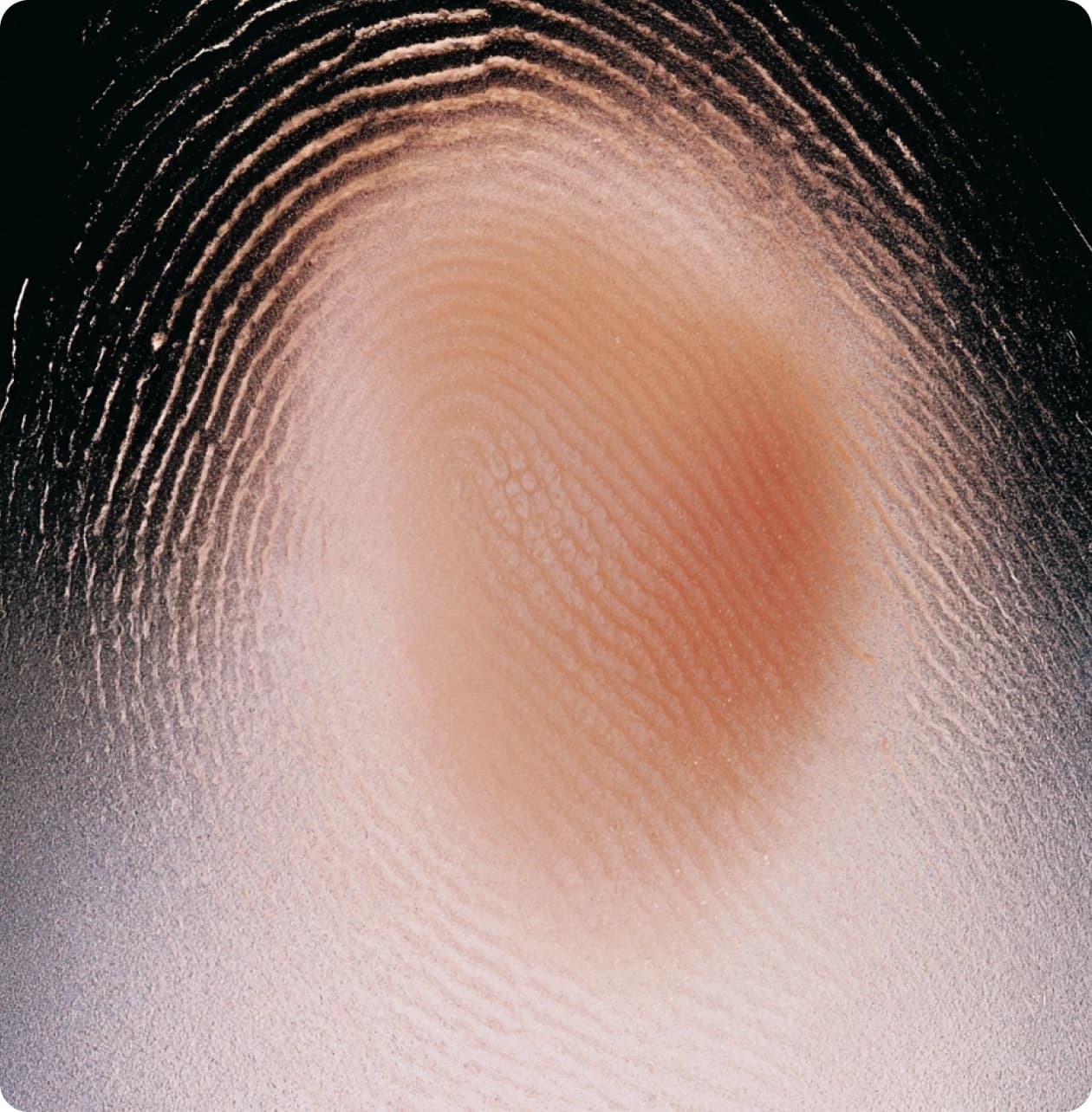
Skin is layered, and each layer answers to a different wavelength. A multispectral scan photographs your face three times in a few seconds, once under standard white light, once under cross-polarized light, and once under ultraviolet light, then analyzes the images computationally.
The three modes exist because they stop at different depths. Together they turn one face into eight separate measurements, each with its own biology.
Standard light behaves like a mirror check with better resolution: it catches surface relief, so texture, pores, and wrinkles appear as patterns of shadow. Cross-polarized light filters out the glare bouncing off the surface, so what remains is light scattered back from below: hemoglobin in vessels and melanin sitting deeper in the skin. Ultraviolet light, near 365 nanometers, is absorbed strongly by epidermal melanin and makes follicular bacteria fluoresce, so it exposes what no visible-light photograph can.
Four are surface readings. Spots are visible discoloration, brown or red. Wrinkles are furrows detected as linear shadows. Texture is the relief of peaks and valleys. Pores are the circular shadowed openings of follicles.
Four are depth readings. UV spots are concentrations of epidermal melanin that absorb ultraviolet light and show dark before they are clinically visible. Brown spots are deeper melanin seen under cross-polarization. Red areas are hemoglobin: dilated vessels, inflammation, acne. Porphyrins are fluorescent by-products of skin bacteria sitting in pores.
The porphyrin your scan counts is mostly coproporphyrin III, made by Cutibacterium acnes, the bacterium implicated in acne. It absorbs near 405 nanometers and fluoresces orange, which is exactly how the camera finds it. The counts are not decorative: in imaging research, coproporphyrin fluorescence correlated with comedone counts at r values of 0.69 to 0.83, and most open comedones fluoresce. A porphyrin reading is a live estimate of bacterial and sebum load, which is why it can move within weeks of changing a routine.
In a reproducibility study, repeated captures deviated by roughly 3% on absolute wrinkle scores, while percentile scores varied closer to 9%; the system's estimated skin age landed within a median of one to two years of true calendar age. The practical reading: trust absolute scores for tracking change, and treat percentiles as context against your peers rather than a precision instrument.
- 01Fulton JE. Utilizing the ultraviolet camera to enhance the appearance of photodamage and other skin conditions. Dermatologic Surgery, 1997.
- 02Patwardhan SV, et al. Measuring acne using coproporphyrin III, protoporphyrin IX, and lesion-specific inflammation. Archives of Dermatological Research, 2017.
- 03Ashkenazi H, et al. Eradication of Propionibacterium acnes by its endogenic porphyrins after illumination with high intensity blue light. FEMS Immunology & Medical Microbiology, 2003.
- 04Henseler H. Assessment of the reproducibility and accuracy of the VISIA camera system for objective skin analysis. GMS Interdisciplinary Plastic and Reconstructive Surgery, 2023.
- 05Oh SJ, et al. Standardized clinical photography considerations in patients across skin tones. British Journal of Dermatology, 2022.